Wellness Testing - Why does my vet keep recommending it?

At some point or another the chances are that your veterinarian has recommended running "wellness" bloodwork on your pet during a visit. While many people appreciate the benefit of wellness testing, there is still some confusion as to what it is and why we (as veterinarians) recommend it. Quite often, the question we're asked is "why run bloodwork, he's acting completely normally"? To fully appreciate why we recommend additional bloodwork on seemingly "normal" pets it's important to understand the thinking behind these recommendations. Many veterinarians (myself included) strive to practice preventive medicine. Preventive medicine is the approach that it's better to prevent a problem in our patients, than react to and treat an already existing (and often more serious) problem down the road. In order to take this proactive approach, we're often looking for signs of impending disease which can sometimes be subtle or require monitoring for change over time. It makes sense that preventing problems is better than trying to play "catch-up" once a disease is in full swing. This isn't to say that all diseases can be addressed this way, but it's good practice to try to prevent illness when we can. Sometimes we can identify potential future problems during our physical examinations - a good example of this would be the prevention of dental disease while examining the mouth. However, the physical exam is just one of the tools we use to assess overall health and sometimes additional testing is needed to identify potential problems. This is especially important because our patients cannot talk to us and tell us how they're feeling. Wellness bloodwork is an example of a tool we use to assess overall health and consists of a comprehensive screen for many diseases. Wellness bloodwork generally consists of a CBC and Biochemical Profile:


CBC: evaluation of red and white blood cells - looks for signs of anemia, clotting disorders and infection (to name a few)

Biochemical Profile: evaluation of body organs such as liver, kidneys, pancreas. Is also used to screen for diseases such as Diabetes Mellitus, Cushings Disease, Thyroid Disease and others.


Recently I was involved in a case that perfectly illustrates how helpful wellness blood testing can be in identifying a potential problem, and initiating therapy in a dog that was showing no signs of disease at all:


A colleague recently called me in to perform laparoscopic liver biopsies on a 7 year old male neutered Standard Poodle. 2 months prior his owner had brought him in for a general annual exam and wellness bloodwork was performed during this appointment. Clinically the dog was acting completely normal and the owner had no health concerns at all, however the bloodwork came back showing some mild to moderately elevated liver values on the biochemical profile. This can sometimes be a normal fluctuation or minor recent liver insult, so the veterinarian recommended monitoring him at home and rechecking the liver values in about 4-6 weeks time. At the recheck examination, the dog was still behaving normally but the liver values had continued to increase so the decision was made to investigate further with laparoscopic liver biopsies. On laparoscopy, the liver appeared smaller than normal, quite nodular, and there were signs of fibrosis (or scarring). Multiple biopsy samples were taken and sent to the lab for interpretation, with concerns about hepatitis or cancer being the underlying cause.


Luckily, the results confirmed that cancer was not present, but did report generalized nodular growth and scarring throughout (suggestive of a slow chronic disease process but not specific).


While this particular disease may not be 100% curable, specific treatments were started to slow down the progression of disease. By catching something early, this veterinarian will now be able to offer therapies that will provide a higher quality of life for a longer period of time. By contrast, if this problem had been identified later due to the dog becoming clinically ill there would have been very little that could have been done to help him (i.e. the damage is already done and he is suffering from liver failure).


Remember: Wellness bloodwork often allows us to identify issues early and prevent smaller problems from becoming big ones!

Recent Posts

Endoscopic view of a large, yellow gallstone lodged in a bile duct.
March 11, 2026
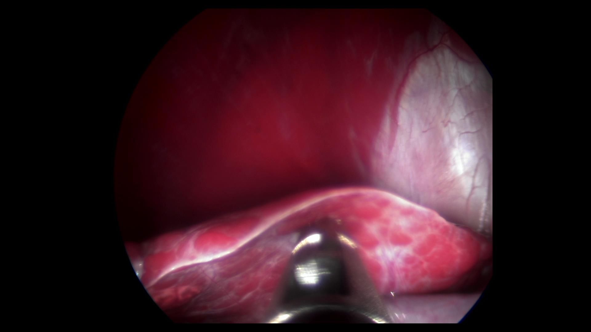
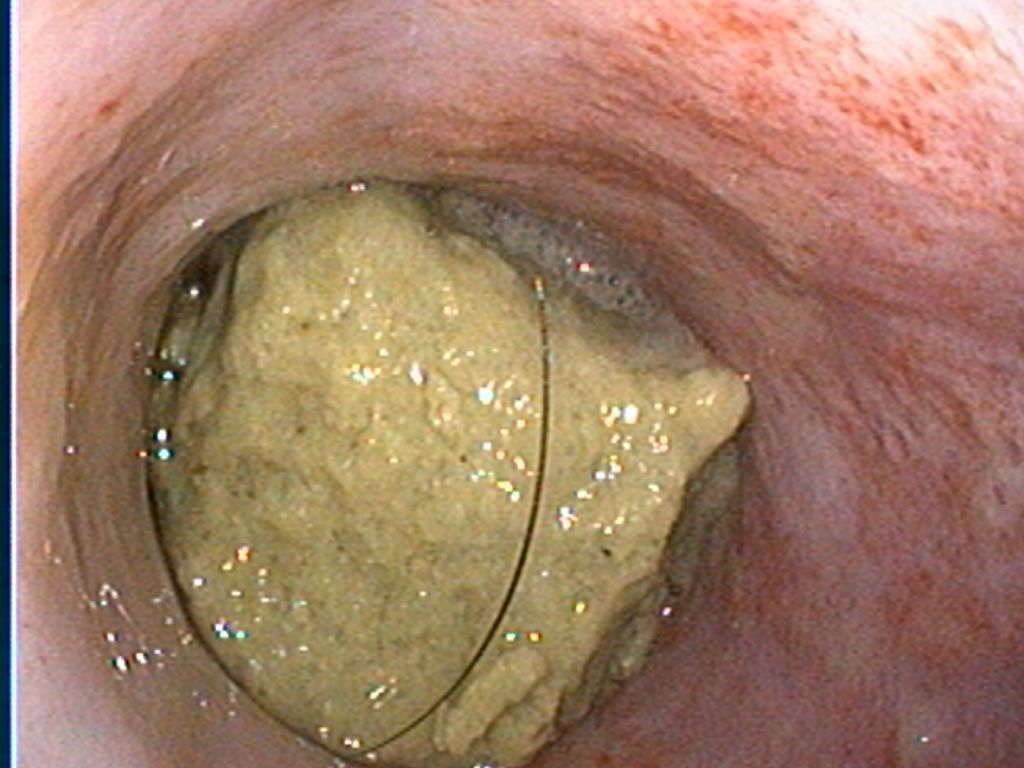
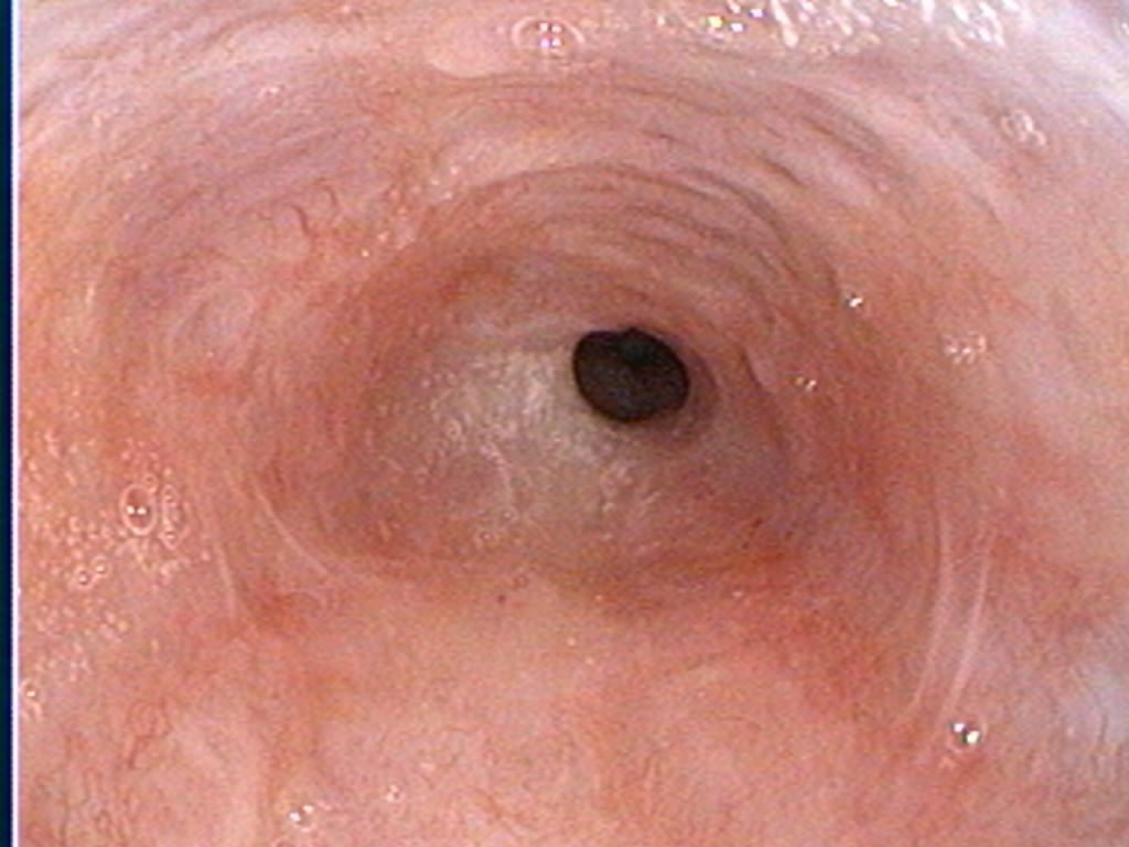
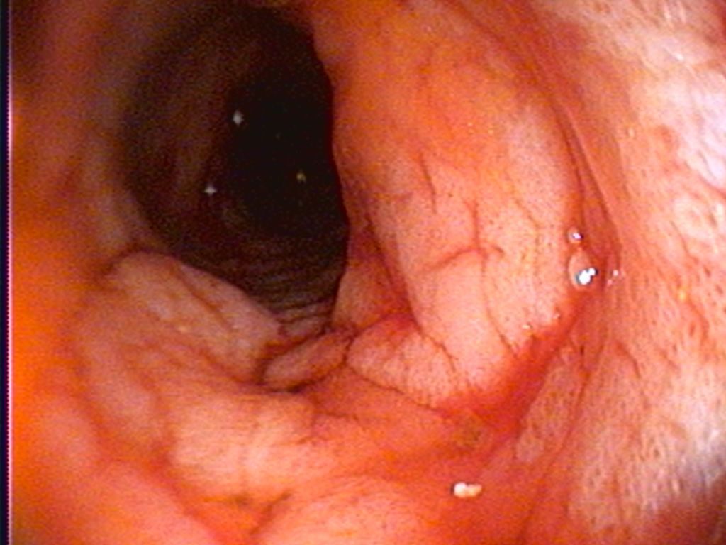
In their never ending quest to amaze pet owners and veterinarians, dogs (and sometimes cats) are always demonstrating how many things they consider "edible". While food products such as bones and corn cobs can be understood, less tempting items such as underpants, toys, stuffed animals, shoes and jewelry tend to make us scratch our heads. Most of these items will make their way down the esophagus into the stomach, and occasionally will pass through their digestive system without any major problems. However, quite often these items will become lodged somewhere and require treatment to be removed (eg: endoscopy to pull it out of the stomach or general surgery if further into the small intestine). On rare occasions a foreign object will become lodged in the esophagus and cause the pet great discomfort. Over the past while I have been asked to remove a few esophageal foreign bodies and these are never pleasant or easy to do. Objects that become lodged in the esophagus are typically larger and more firm in nature, making it harder for the pet to swallow down to the stomach. Once the object becomes stuck it can be difficult to dislodge and swift treatment is the best chance for preventing more serious secondary trauma. In particular, animals with esophageal foreign bodies are at a risk of pressure ulcerations forming in the esophagus (where the foreign material is tightly wedged). The esophagus is not able to distend like a stomach would and pressure sores (ulcers) can start to form fairly quickly. Should the foreign material be successfully removed, the esophagus begins to repair itself and scar tissue can form. If a large amount of scar tissue forms in an area there is a risk of stricture (shrinking down or narrowing of the opening in the esophagus). In extreme cases these strictures can cause the esophagus to become so narrow that food cannot get through. When this happens the process for opening that narrowed spot back up can be time consuming, uncomfortable for the pet, and expensive. In many cases, signs of an esophageal foreign body can be difficulty swallowing, repeated swallowing motions despite not currently eating anything, drooling, regurgitation when trying to eat, and even difficulty breathing if the object is putting pressure on the trachea (airway). BOTTOM LINE: If you think your pet has an object stuck in their throat, don't wait! The sooner you can get to a veterinarian to investigate the better. Case #1: 5 year old small breed dog that was eating a dental chew and swallowed a large piece that became lodged in the esophagus. She was not experiencing any respiratory issues and was stable, so the esophagoscopy was performed later that day. The foreign object was removed leaving a mild to moderate amount of inflammation to the tissue (seen as small red depressions on the lower left image. This little girl made a full recovery. Case #2: 10 year old small breed dog with history of regurgitation for a number of days prior to presentation to his veterinarian. Radiographs showed a large amount of foreign material in the esophagus and esophagoscopy was performed to attempt to remove the obstruction. During an extremely difficult procedure we removed multiple pieces of a small plastic toy combined with a large amount of hair. The esophagus was extremely ulcerated and we were concerned about a secondary stricture formation. Despite aggressive therapy a severe stricture did form and ultimately humane euthanasia was elected. While unfortunate, this serves to demonstrate the importance of recognizing a problem and treating it as quickly as possible.
Endoscopic view inside the esophagus, showing a dark, round opening at the center surrounded by pink tissue.
March 11, 2026
It's happened to all of us - we're in the middle of a surgery and our technician tells us that the patient is regurgitating. While we continue to operate, our techs quickly clean out the mouth and double check to ensure the endotracheal cuff is snug. Now what? There are a variety of ways to address an obvious regurgita
Endoscopic view of the colon interior, showing reddish, textured tissue and folds.
March 11, 2026
In my last post I discussed the options available when collecting intestinal biopsy samples. While both flexible endoscopic samples and laparoscopic assisted intestinal biopsy have their benefits, it really does come down to individual expectations and suspicion of disease(s). In this post, I thought I would summarize